
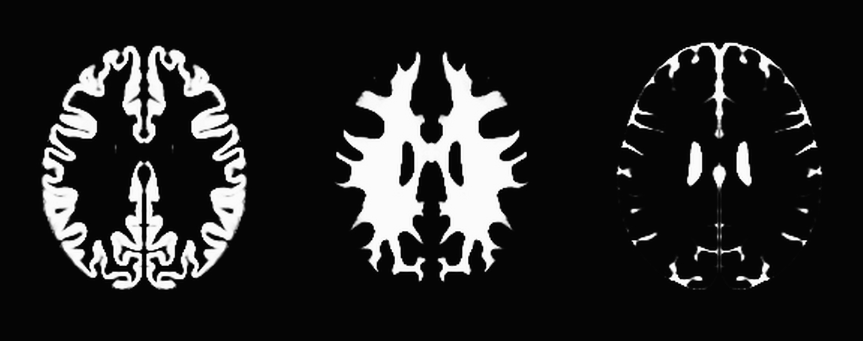
Voxel Based Morphometry (VBM)
VBM is an analysis technique which allows analysis of local differences in brain anatomy. Brains are warped to a common template and the images are smoothed so that each voxel represents an average of itself and it's neighbors. The whole brain is compared across brains at every voxel to identify differences. This technique allows one to investigate differences in Grey Matter, White Matter and Cerebral Spinal Fluid across groups. Oftentimes, we have used this technique in conjunction with fMRI to verify if differences detected with fMRI were attributed to underlying macrostructural changes or solely to a difference in the Blood Oxygen Level Dependent (BOLD) signal.

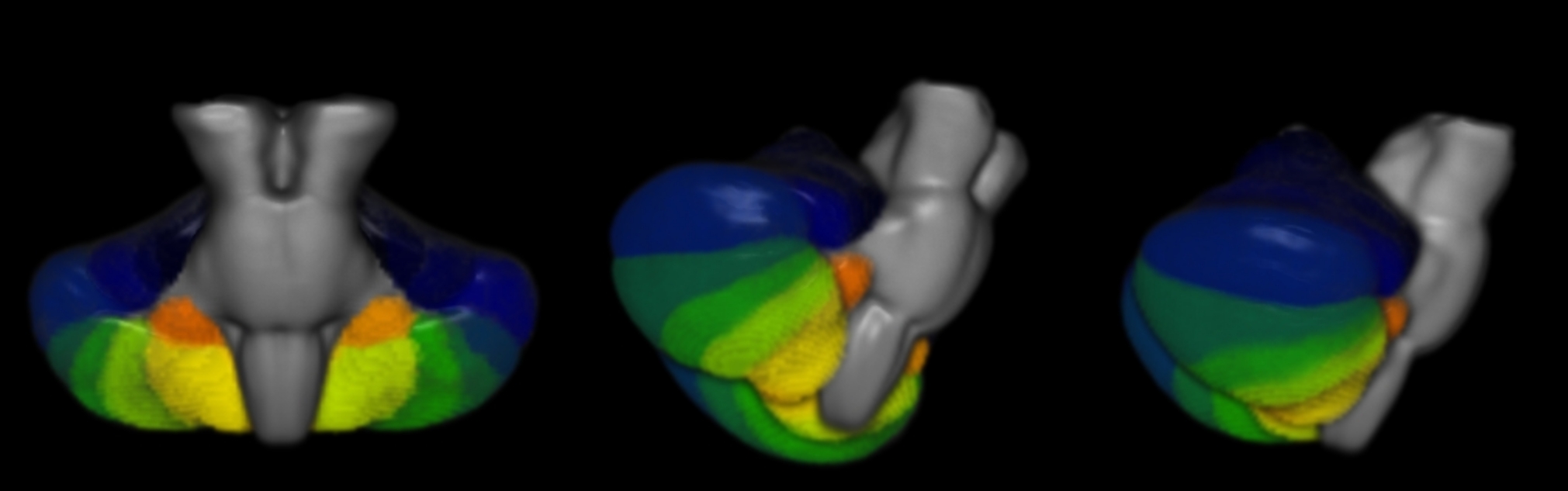
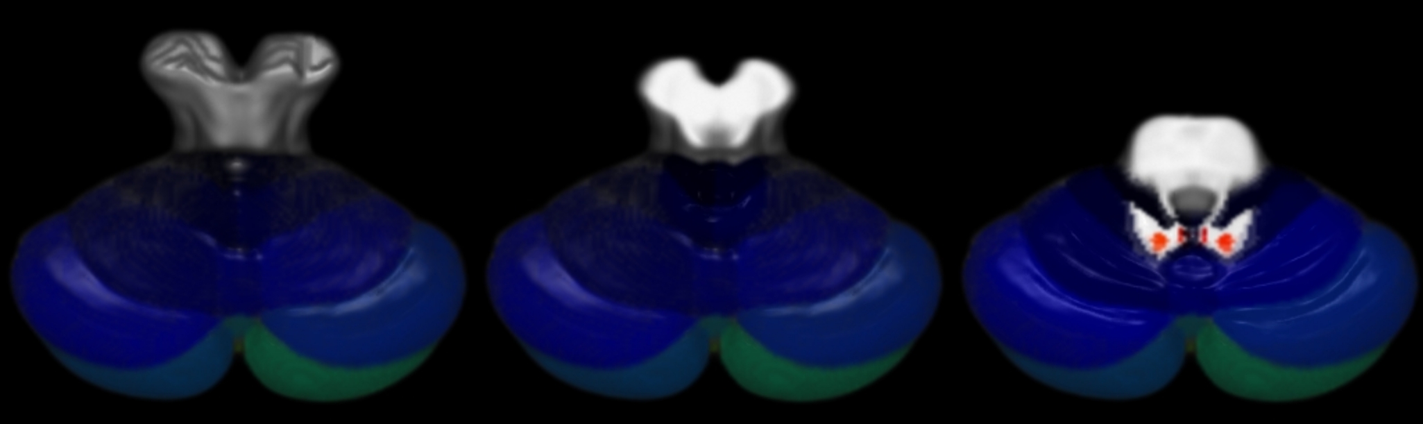
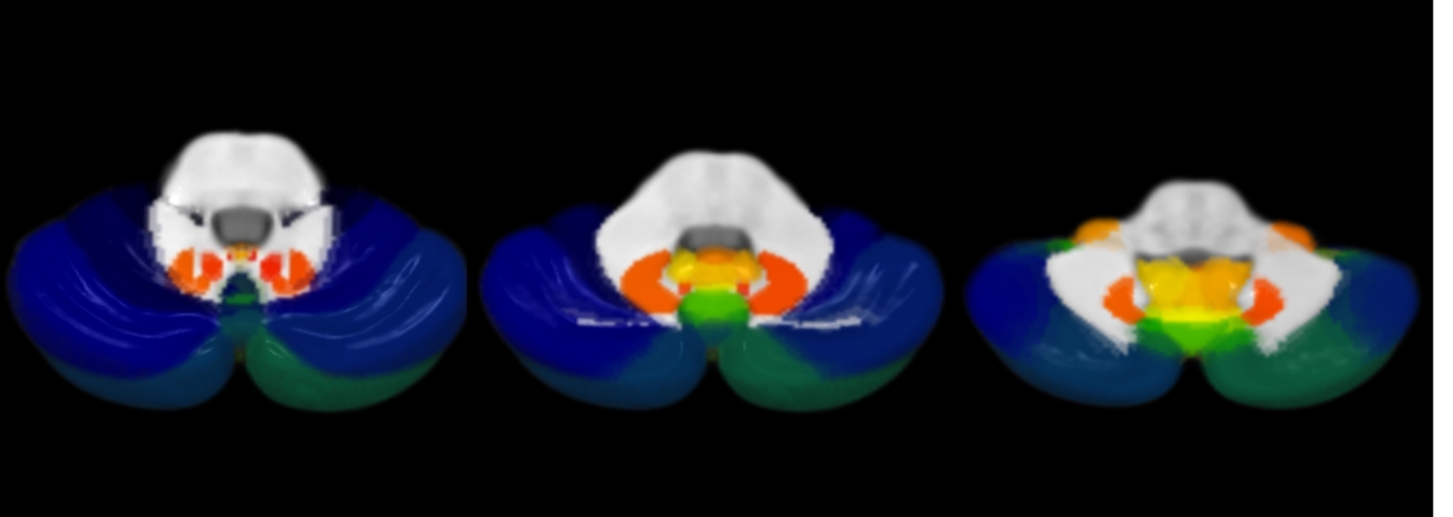
Cerebellar Normalization
Typically, whole brain common space templates (such as Talairach or MNI space) have poor resolution of cerebellar structures due to poor spatial resolution in this region. SUIT is an SPM toolbox developed for high accuracy spatial normalization of the brainstem and cerebellum. This toolbox can be used for VBM, fMRI and other types of image analysis which require high degree of spatial overlap within the cerebellum. This technique was used in functional MRI (fMRI) and resting state fMRI studies to examine healthy controls and subjects with movement disorders. Improved sensitivity of measures from the cerebellum were used to examine various aspects of motor function as well as deficits in clinical populations.
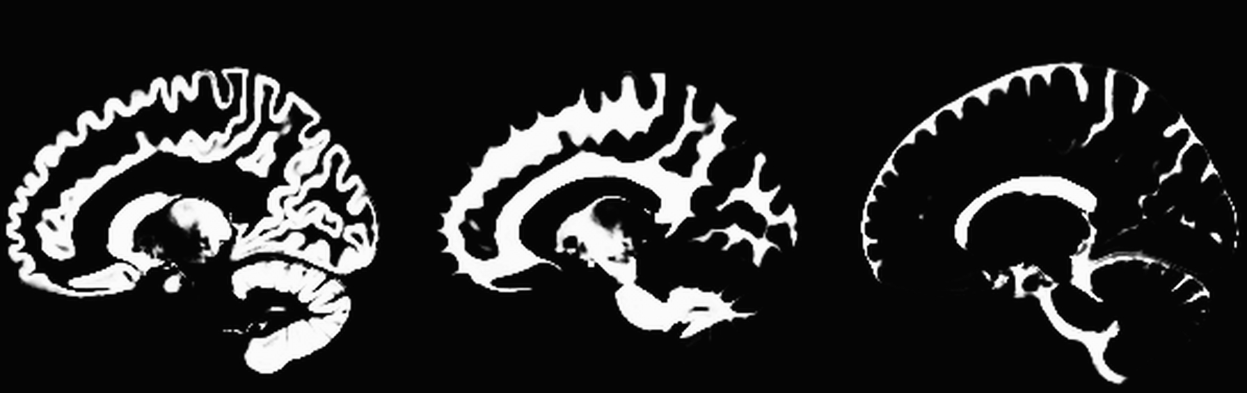
Cortical Thickness
Cortical thickness is currently being used to investigate subtle differences between healthy controls, Parkinson's Disease (PD) subjects, and PD with mild cognitive impairment (PD-MCI). I have developed a high resolution surface model based on the MNI152 template which significantly improves spatial overlap and accuracy of results. By nonlinearly warping thickness values to a cortical surface map, one is able to detect cortical changes.

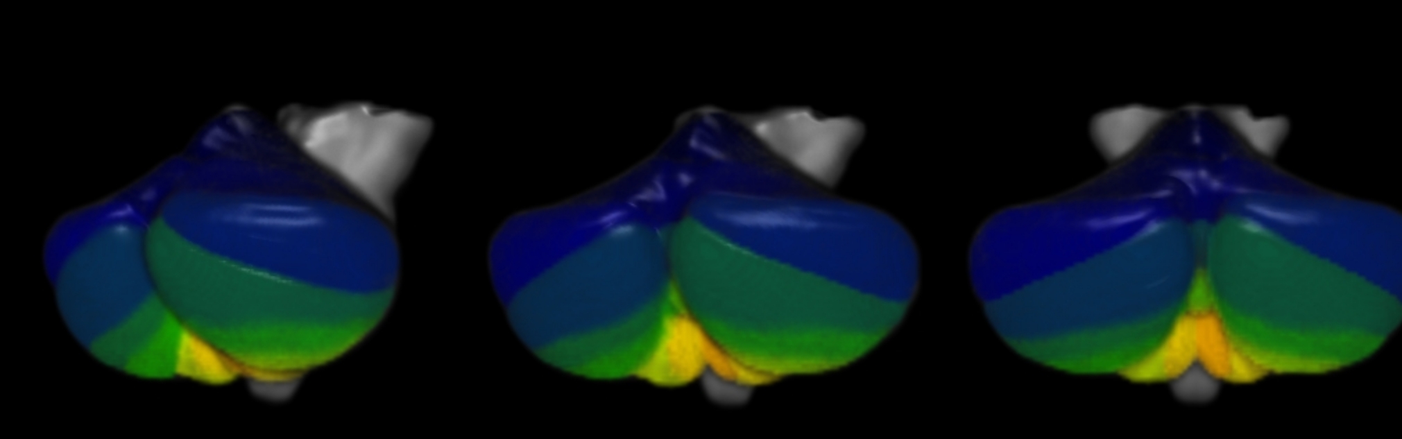
Shape Analysis
Shape analysis of subcortical brain structures can help identify conformational changes in the shape of the underlying region of interest. Metrics derived from quantifying the structural shape change have been shown in other studies to be indicative of neurodegenerative diseases such as Parkinson's disease. A newly developed pipeline will be used to evaluate cognitive deficits in Parkinson's disease.
