
Task-based fMRI
Task based functional magnetic resonance imaging (fMRI) in conjunction with advanced cerebellar normalization procedures have been used to investigate regions in the cerebellum involved in motor control. Using this technique, specific cerebellar regions were identified in which BOLD changes scaled with amplitude of isometric force production as well as rate of change of isometric force production. Task based fMRI was also used to compare subtypes of Parkinson's disease (Tremor and Non-tremor dominant PD), as well as compare PD to Essential Tremor subjects.

Resting State fMRI
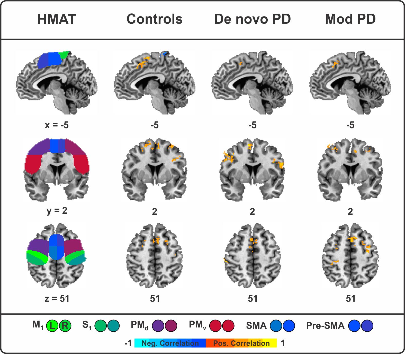
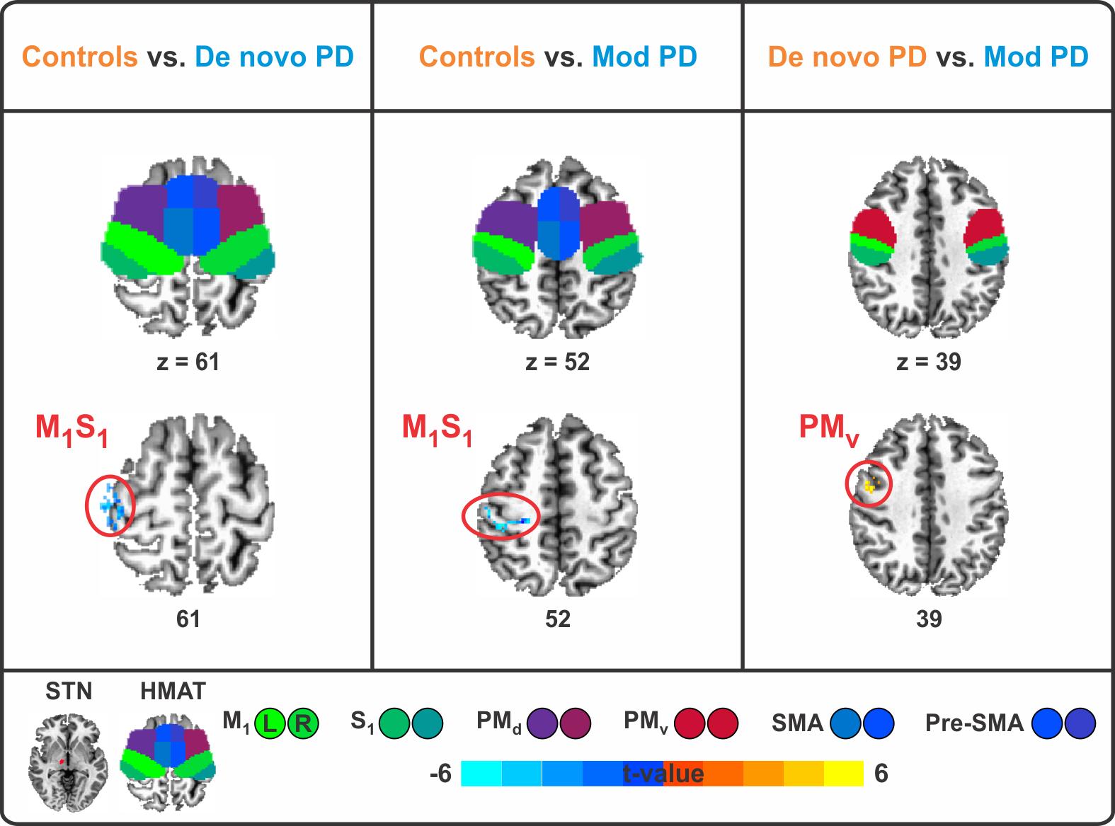
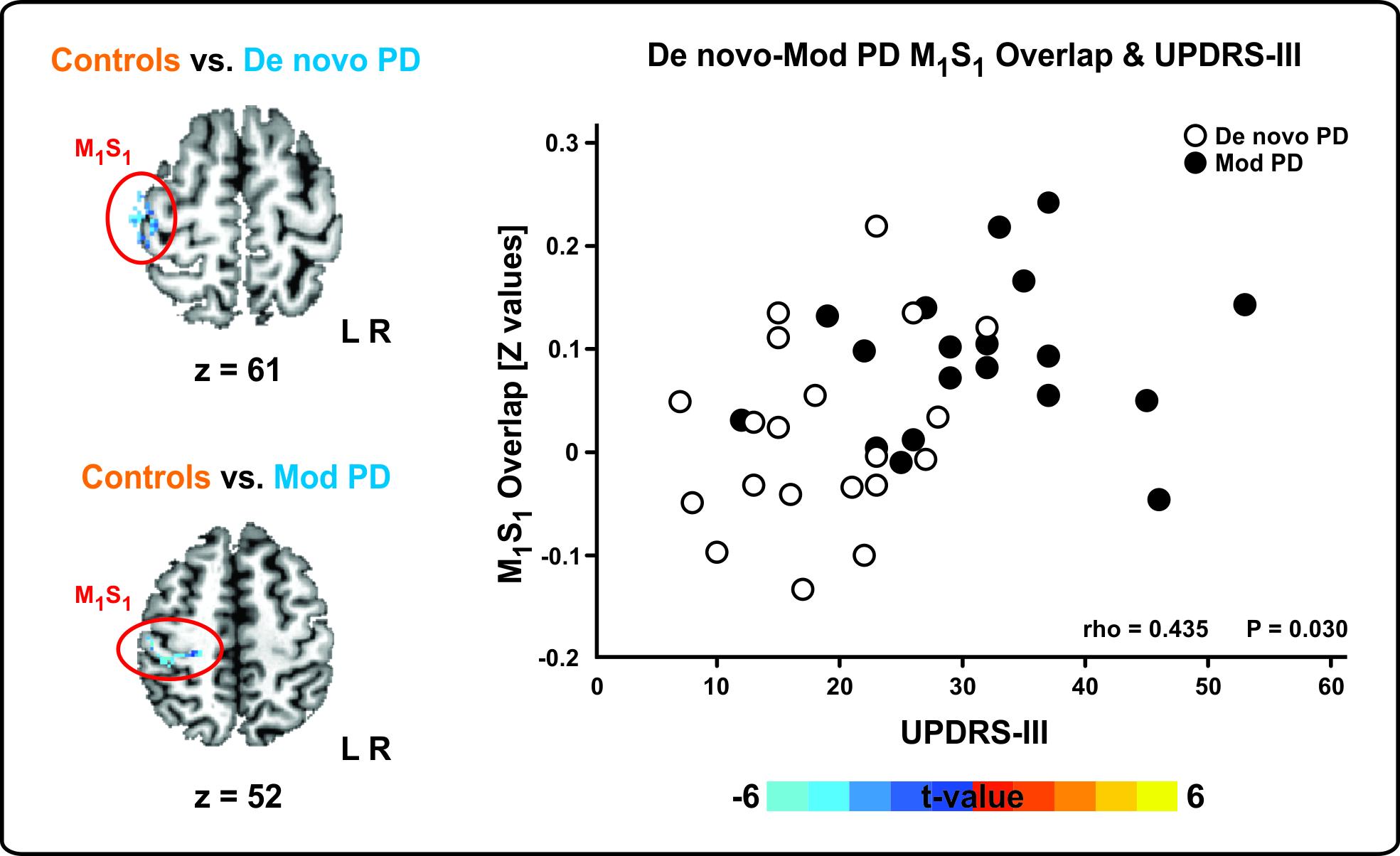
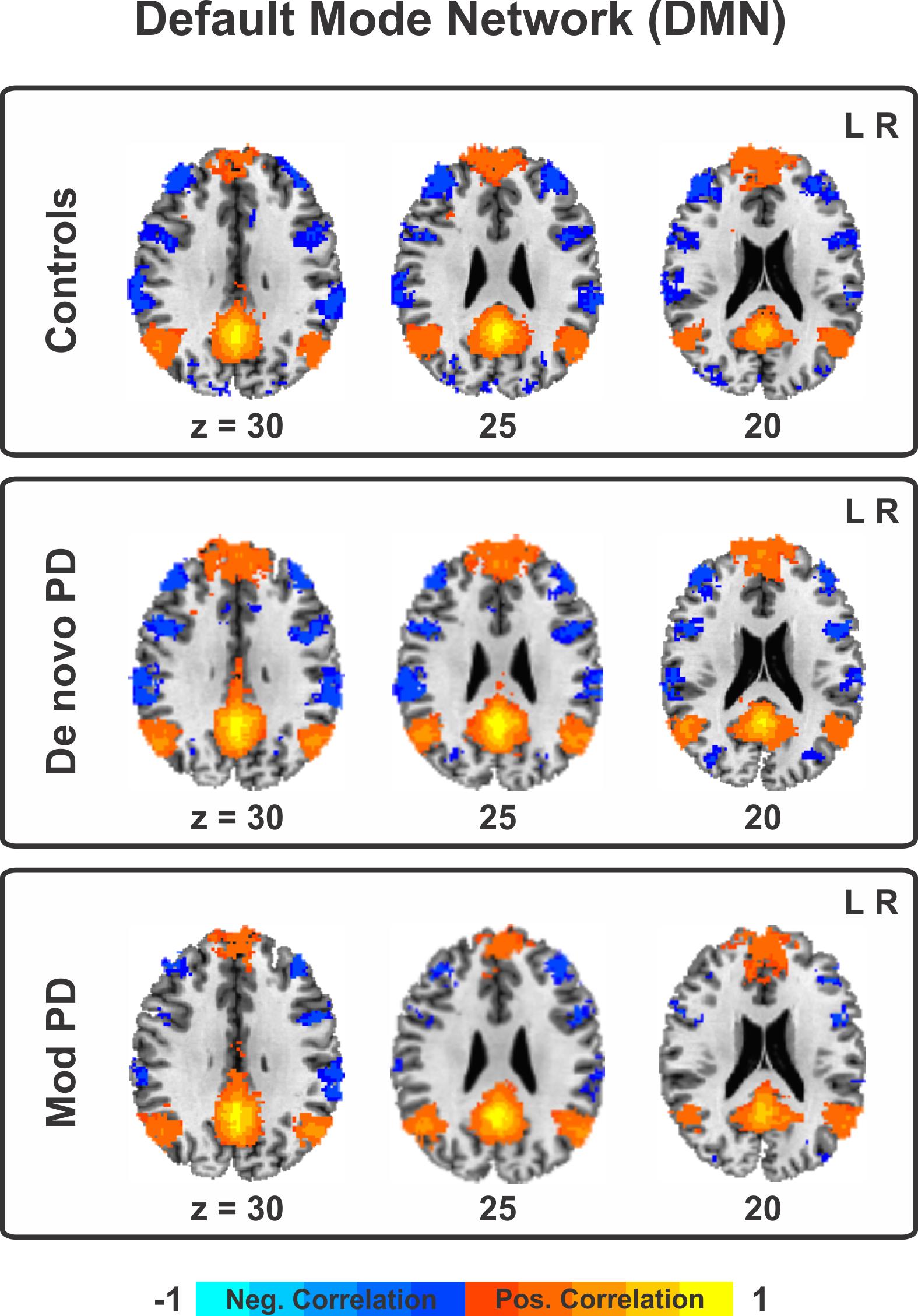
Resting state fMRI is a functional imaging technique which can be used to evaluate functional connections within the brain when no explicit tasks are performed. We have developed a robust pipeline which I used to evaluate seed based functional connectivity between the subthalamic nucleus and motor cortices in PD subjects. Additionally, we have used this technique to examine which pathways are commonly and differentially affected in PD and Progressive Supranuclear Palsy. Furthermore, we have used extensions of this technique to improve the detection of seizure networks in epilepsy patients in which structural MRIs appeared normal.
Technical Development
In terms of technical development, we are interested in developing more advanced analysis techniques related to RS-fMRI. One area relates to dynamic distortion correction related to account for both motion and geometric distortions as a result of B0 field inhomogeneities. Current techniques use a static fieldmap which may not be accurate as the brain shifts within the magnetic field. These corrections will substantially improve multi-modal image analysis. Additionally we are interested in developing advancements related to physiological noise contamination. Many of these techniques can also be adapted to work with Diffusion Weighted Images.